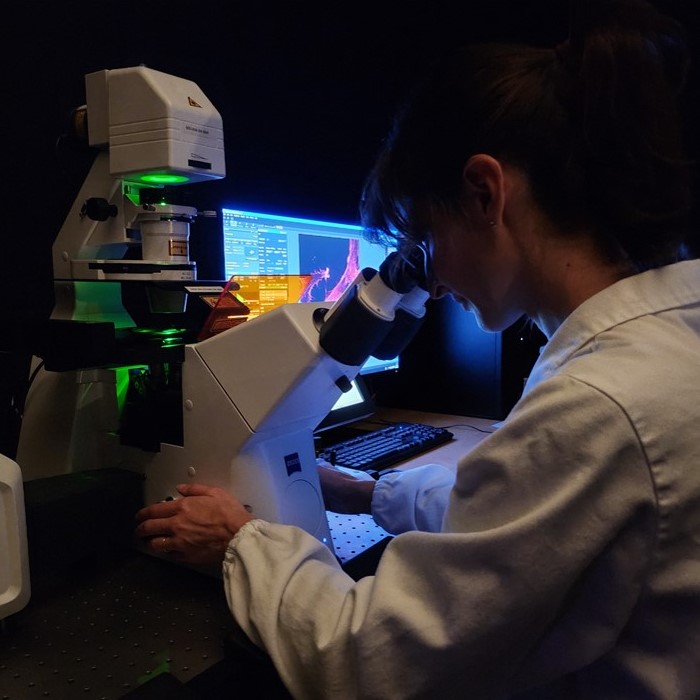
Imaging
Optical microscopy is one of the fastest-growing technologies in scientific research. Unlike other techniques in life sciences, it allows to see biological processes in their natural, physiological context.
Imaging
Optical microscopy is one of the fastest-growing technologies in scientific research. Unlike other techniques in life sciences, it allows to see biological processes in their natural, physiological context.

Facility manager Dr. Diana Corallo
- Cell Counting
- Cell Cycle Analysis
- Protein Localization, Distribution, and Co-localization in Cells or Tissues
- Live Cell Assays
- Cellular Morphology (e.g. Neurite Growth)
- Capture and Analysis of Rare Events
- Other applications requiring optical imaging (e.g. new materials or structures >100 nm)
- High-Quality Data Production: we provide you multi-parametric datasets for deeper insights for each of your samples.
- Faster Progress: accelerates research and discovery thanks to our cutting-edge imaging technologies.
- Multi-disciplinary support: we can offer all the expertise of our Institution not only our microscopes.
- Data-Driven Decisions: enables more informed and confident decisions thanks to our advanced AI-driven tools.
- Flexibility and Scalability: we can support you from sample preparation to data analysis.
Zeiss LSM800 Confocal Microscope
with Airyscan technology for super-resolution imaging, providing high-quality resolution at all depths using standard sample preparation.
Zeiss Axio Observer 7 Inverted Microscope
with a Colibri LED light source and ApoTome III structured illumination system for enhanced contrast and resolution. It also features an incubator chamber for live sample incubation, offering stable temperature and CO2 control for long-term imaging.
Zeiss Axio Imager M1 Upright Microscope
for imaging histological and slide-mounted samples with color and grayscale cameras. It supports Z-stack and tiled image acquisition.
Leica Thunder Imager Model Organism
for fast, 3D imaging of large specimens, such as whole organisms or organoids. It offers Z-stack acquisition and computationally cleared images in real-time.
Olympus IXplorer
for high content imaging, coupled with a AI-base software for High Content analysis.
