Research Area: Biotecnologie Mediche
Group Leader
 The research activities of the Optics and Bioimaging Group are focused on the development of micro- and nano-platforms and devices for different applications: from nano-optics to telecom, from holography to microscopy and astronomy, from biology to materials science. Nanotechnology development and application rely on the intersection of expertise from many science and engineering disciplines. The Optics and Bioimaging Group at IRP is structured synergistically to foster integration among the physical, biological, and clinical sciences. Our bioimaging, optical and lab-on-chip systems intend to overcome these limitations following innovative technological solutions that conjugate physics with biology and medicine.
Bioimaging
The group is highly specialized in bioimaging, from experimental design, to labelling protocols, data acquisition and elaboration.
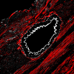
In the Optics and Bioimaging Group, we have designed and assembled a custom microscope capable of performing Two-Photon Microscopy, Label-Free Microscopy that is currently being upgraded for STED super-resolution. Two-Photon Microscopy (TPM) takes advantage of the quasi-simultaneous absorption of two photons by a molecular receptor in a single quantum event. Despite the need for very intense light sources, the NIR wavelengths used in TPM are significantly less prone to scattering and absorption from thick specimens and, therefore, exhibit longer penetration depths. Additionally, TPM has an inherent capability of performing axial sectioning and confines the photo-bleaching effect inside a very limited volume. When specimens are very susceptible to photo-bleaching and photo-damages or when any staining process must be avoided in order to preserve thes pecimens in their original conditions, the use of Label-Free Microscopy (LFM) is preferable. LFM can be applied to study samples with no need for exogenous fluorescent probes, while keeping the main benefits of TPM.
The group is also collaborating in confocal and live microscopy projects to optimize data acquisition and elaboration.
Optics
The generation and control of complex light beams, i.e. beams with unusual distribution of amplitude and phase, requires the manipulation of the wavefront at the micrometric scale. This is achievable by realizing specific 3-dimensional micro-structured patterns on a transparent material, which act locally on the incident light in order to transfer specific phase distributions to the electromagnetic field. Our activities cover the design, the fabrication and characterization of nanostructures and nano-objects. The applications of these optical devices range from the telecommunications world (OAM-mode division multiplexing for enhanced information capacity), to microscopy (high-resolution optical microscopes).
The Optics and Bioimaging Group has developed a high-sensitivity SPR-based biosensor for applications in the biomedical field. A phase-interrogation setup based on polarization scan allows a high miniaturization and integration of the plasmonic sensor in lab-on-a-chip platforms. The detection setup (patented) is simple, cost effective and guarantees high sensing speeds and performances.
Group Members
Giulia Borile PostDoctoral Researcher
Pietro Capaldo PostDoctoral Researcher
Gianluca Ruffato PostDoctoral Researcher
Deborah Sandrin PostDoctoral Researcher
Andrea Vogliardi Graduated student
Selected Publications
A Comprehensive Comparison of Bovine and Porcine Decellularized Pericardia: New Insights for Surgical Applications. Zouhair S, Sasso ED, Tuladhar SR, Fidalgo C, Vedovelli L, Filippi A, Borile G, Bagno A, Marchesan M, Giorgio R, Gregori D, Wolkers WF, Romanato F, Korossis S, Gerosa G, Iop L.Biomolecules. 2020 Feb 28;10(3):371. doi: 10.3390/biom10030371.
Multiplication and division of the orbital angular momentum of light with diffractive transformation optics. Ruffato G, Massari M, Romanato F. Light Sci Appl. 2019 Dec 5;8:113. doi: 10.1038/s41377-019-0222-2. eCollection 2019.
Label-free, real-time on-chip sensing of living cells via grating-coupled surface plasmon resonance. Borile G, Rossi S, Filippi A, Gazzola E, Capaldo P, Tregnago C, Pigazzi M, Romanato F. Biophys Chem. 2019 Nov;254:106262. doi: 10.1016/j.bpc.2019.106262. Epub 2019 Sep 3.
Total angular momentum sorting in the telecom infrared with silicon Pancharatnam-Berry transformation optics. Ruffato G, Capaldo P, Massari M, Mafakheri E, Romanato F.Opt Express. 2019 May 27;27(11):15750-15764. doi: 10.1364/OE.27.015750. PMID: 31163766.
Multimodal label-free ex vivo imaging using a dual-wavelength microscope with axial chromatic aberration compensation. Filippi A, Dal Sasso E, Iop L, Armani A, Gintoli M, Sandri M, Gerosa G, Romanato F, Borile G. J Biomed Opt. 2018 Mar;23(9):1-9. doi: 10.1117/1.JBO.23.9.091403.
Contatti

Corso Stati Uniti, 4 F
35127 Padova
Phone: +39 049 9640111
Fax: +39 049 9640101
info@irpcds.org
Orario di apertura: lun-ven 8:30 – 17:30


